
Fundus Photography vs. Wide Angle Retinal Photography
26-08-2023
In the world of modern eye care, advanced diagnostic techniques have revolutionized our ability to delve into the intricacies of ocular health. Two such diagnostic scans, fundus photography and wide-angle retinal photography, are often used interchangeably by patients seeking comprehensive eye examinations and marketers trying to advertise eye care services. However, they are not synonymous and offer unique insights into retinal health. Understanding the difference between these two techniques is crucial for patients to make informed decisions about their eye health assessments.
Fundus Photography: Exploring the Inner Layers
Fundus photography is a diagnostic technique that captures detailed images of the fundus, the innermost layer of the eye that includes the retina, optic nerve, and blood vessels. Using a fundus camera, optometrists capture high-resolution images of this crucial region, allowing them to monitor and detect abnormalities, such as macular degeneration, diabetic retinopathy, and glaucoma.Fundus Photography = 30° Field of View
Fundus photography primarily focuses on capturing a specific area of the retina, providing detailed information about its condition. These images are a vital reference for tracking changes over time, enabling early intervention and tailored treatment plans.
Wide Angle Retinal Photography - A More Complete Picture
On the other hand, wide-angle retinal photography takes the diagnostic process a step further by capturing a panoramic view of the entire retina, including that of typical fundus photography. This technique utilizes advanced imaging technology to capture a more comprehensive image, providing a wider field of view. This broader perspective offers optometrists the opportunity to detect peripheral abnormalities and retinal disorders that may not be visible with traditional fundus photography.Fundus Photography = 120° Field of View
Wide-angle retinal photography is particularly advantageous for conditions like retinal detachments and peripheral retinal degenerations. The ability to visualize the periphery allows for early detection and timely management of potentially sight-threatening issues.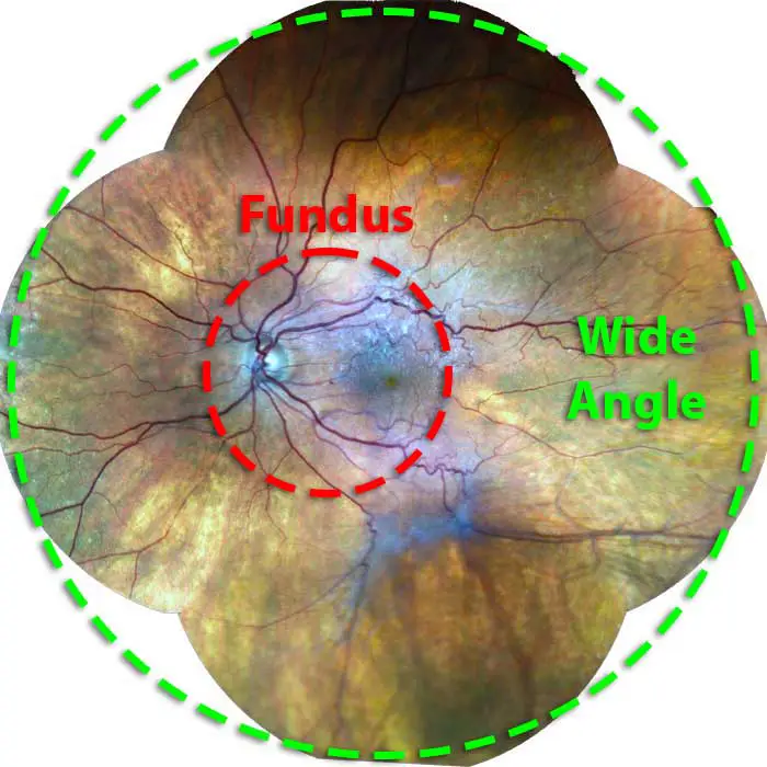
Why the Distinction Matters
While fundus photography and wide-angle retinal photography are both invaluable tools for assessing retinal health, their distinct purposes offer unique advantages. Fundus photography excels in capturing detailed images of specific areas, enabling close monitoring of retinal conditions. On the other hand, wide-angle retinal photography includes fundus photography AND paints a broader picture, revealing abnormalities that extend into the periphery. Patients seeking comprehensive eye examinations must understand these distinctions. Clinics may offer either one or both of these techniques during an eye exam. It is imperative for patients to inquire about the types of tests and scans provided by their optometrist, as they are not uniform across the industry, even though the cost of an exam may be the same.Empowering Informed Decisions
As technology continues to evolve, the world of eye care continues to expand with innovative diagnostic tools. The distinction between fundus photography and wide-angle retinal photography highlights the multifaceted approach to assessing retinal health. By understanding the differences in the scans, patients are empowered to make informed decisions about their eye health assessments. When seeking an eye examination, don't hesitate to engage with your optometrist. Inquire about the diagnostic techniques they employ and how these techniques can contribute to your comprehensive eye health evaluation. By taking an active role in your eye care journey, you ensure that your vision is in the best hands possible. Schedule an eye exam with our Edmonton optometrists and experience what technologically advanced and thorough eye care looks like today! Schedule An AppointmentFYEyes Blog Posts
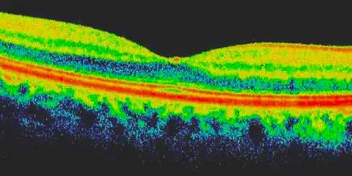
Unmasking The Illusion Of "3D" OCT Scans
Optical Coherence Tomography provides only cross-sectional views of ocular tissues and the retina.
Importance of Advanced Eye Exam Technologies
Advanced eye exam technologies promote the early detection of eye diseases.
OCT: Optical Coherence Tomography
Optometrists use OCT to assess the retinal nerve layers of the retina.
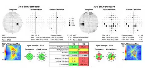
Visual Field Test
Perfect vision requires that you can see well everywhere in your field of vision.

Adult Eye Exams
Our advanced eye exams consist of 25+ modern tests and digital scans to assess eye health, function, and visual acuity.

Child Eye Exams
Give your child a clear future with an annual eye exam from our experienced Edmonton optometrists.

Senior Eye Exams
Maintain your vision through your golden years with gold standard eye care from the optometrists at our Edmonton eye clinic.

Contact Lens Eye Exams
Our eye exams for contact lens wearers include test and digital scans to assess eye health, function, visual acuity, and lens fit.

Diabetic Eye Exams
Managing diabetes requires regular eye exams to ensure that diabetes is not causing irreversible vision loss.

Dilated Eye Exams
Dilating the eyes enables our Edmonton optometrists to see more of the eye so that you many never see less.
Our Edmonton Eye Exams Are Comprised Of 4 Phases Of Evaluation

1. Eye Exam Pre-Testing
Corneal Thickness | Intraocular Pressures | Visual Field
Pre-testing is a detailed process that gathers all necessary information for the optometrist in advance of the optometrist-administered eye examination. This process involves completing a detailed patient history, as well as a series of standard tests. Pre-testing is an essential part of the comprehensive eye exam process, providing valuable information and visuals for both the optometrist and the patient.
More About Pre-Testing »
2. Advanced Diagnostic Testing
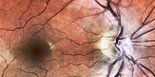
Retinal Photography, OCT, Topography
eye-deology Vision Care differentiates itself from other clinics by having the most advanced modern diagnostic specialty testing equipment. Specialty equipment, such as a wide-angle high-resolution retinal imager, Optical Coherence Tomography (OCT), Humphrey Visual Field Analyzer and corneal topographer, ensures that patients receive the best comprehensive eye care.
More About Advanced Testing »
3. Optometrist Examination
Health Assessment & Disease Diagnosis
eye-deology Vision Care Edmonton optometrists perform a multitude of tests and assessments to evaluate ocular health, eye coordination, and visual acuity. In addition, they also evaluate the results of the tests and scans performed during pre-testing. As part of patient education, our optometrists also take the time to show and explain results to patients.
More About Doctor Exam »
4. Eye Glass Consult
Prescription | Lens Selection | Digital Fitting
If you require corrective lenses to improve your vision, our licensed opticians will customize their fit to your unique attributes, needs, lifestyle, and budget. Our opticians are happy to provide you with information about the latest eyeglass frame and lens technologies available so you can make informed decisions and begin seeing and looking your best.
More About Eyewear Consult »